
AQA Syllabus focus:
'The structure and function of sensory, relay and motor neurons.'
Neurons are specialized cells that carry information through the nervous system. For AQA Psychology, you need to know how sensory, relay, and motor neurons are built and how each supports behavior.
What neurons do
Neurons are specialized cells built to receive, carry, and pass on information. Their structure is not accidental: each part of the cell helps it move impulses in a particular direction. In A-Level Psychology, three main neuron types are usually identified: sensory neurons, relay neurons, and motor neurons. They differ in shape, location, and role, but all contribute to rapid coordination of responses.
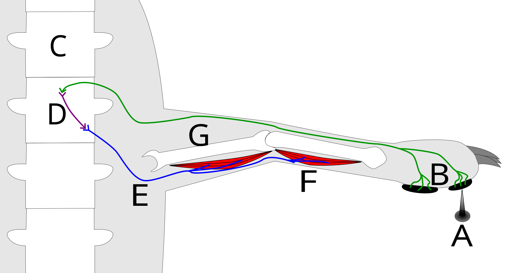
A labeled reflex arc showing the flow of information from a stimulus along a sensory neuron into the spinal cord, through an interneuron (relay neuron), and out via a motor neuron to a muscle (effector). This reinforces the core AQA idea that relay neurons in the CNS link sensory input to motor output in a fast, coordinated pathway. Source
Neuron: A specialized nerve cell that carries information around the nervous system using electrical and chemical signals.
Most neurons share some basic features:
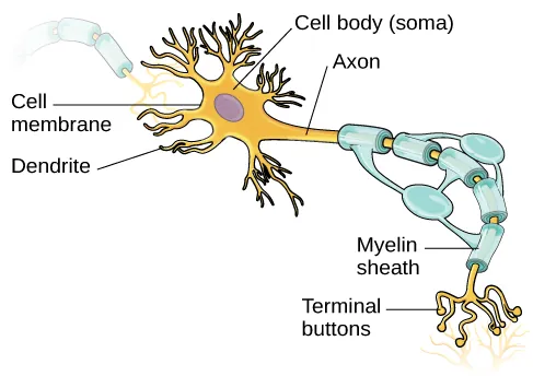
A labeled diagram of a prototypical neuron highlighting dendrites (input region), the cell body/soma, the axon (conduction pathway), and axon terminals (output region), with myelin shown as insulation along the axon. This provides a clear visual anchor for the direction of information flow and the structural terms used throughout the sensory, relay, and motor neuron descriptions. Source
Dendrites receive input from other cells or receptors.
The cell body contains the nucleus and keeps the neuron alive.
The axon carries the impulse away from the cell body.
The axon terminals pass the signal to the next neuron, muscle, or gland.
The exact arrangement of these parts changes depending on the job the neuron performs.
Sensory neurons
Sensory neurons carry information from receptors to the central nervous system. They are activated when a stimulus such as light, pressure, temperature, or sound is detected.
Receptor: A cell or sense organ that detects a stimulus and converts it into a nerve impulse.
Structure
In a typical sensory neuron, the cell body is located partway along the neuron, rather than at one end. One long dendron carries the impulse from the receptor toward the cell body, and a short axon carries it from the cell body into the CNS. This makes sensory neurons look different from motor neurons in diagrams. Many sensory neurons are also myelinated, meaning they have a fatty insulating covering that helps impulses travel more quickly.
Function
The function of a sensory neuron is to detect change in the internal or external environment and send that information to the CNS for processing. Its structure suits this role:
the long dendron allows information to travel from a receptor over some distance
myelin helps increase the speed of transmission
the ending of the axon allows the message to be passed to neurons in the CNS
Sensory neurons are therefore the main route by which stimulation enters the nervous system.
Relay neurons
Relay neurons are found within the CNS and link other neurons together. Their main function is to pass information between sensory and motor neurons, although they can also connect different neurons in more complex pathways.
Structure
Relay neurons usually have short dendrites and a short axon. Because they often only have to transmit information across a small distance inside the brain or spinal cord, they do not need the long dendron of a sensory neuron or the long axon of a motor neuron. Their branching structure allows them to receive input from several neurons and send output onward.
Function
The function of a relay neuron is not simply to carry on a message unchanged. It helps organize neural pathways by selecting, combining, and passing information to the appropriate neuron. In exam answers, it is important to state that relay neurons act as the link between sensory input and motor output. Their short connections are well suited to rapid communication across networks in the CNS.
Motor neurons
Motor neurons carry impulses from the CNS to effectors, producing a response such as muscle contraction or gland activity.
Effector: A muscle or gland that produces a response after receiving signals from a motor neuron.
Structure
A motor neuron has a cell body at one end with many short dendrites spreading from it. These dendrites receive input from other neurons. From the cell body, a single long axon extends to the effector. The axon may be covered by a myelin sheath, which helps the impulse move quickly along the neuron. At the far end, axon terminals branch out so the motor neuron can communicate with the muscle or gland it controls.
Function
The function of a motor neuron is to carry the final command that produces action. Its structure supports this role:
many dendrites allow input from relay neurons or other cells
a long axon allows the impulse to travel from the CNS to a distant effector
branching terminals help one motor neuron influence many muscle fibers or parts of a gland
Motor neurons therefore convert neural processing into an observable response.
Comparing the three neuron types
The main differences are easiest to remember by linking structure to function.
Sensory neurons: from receptor to CNS; typically have a long dendron, cell body along the length, and short axon
Relay neurons: found within the CNS; usually have short dendrites and a short axon because they connect nearby neurons
Motor neurons: from CNS to effector; usually have many short dendrites, a cell body at one end, and one long axon
A common exam mistake is to describe only what each neuron does. A stronger answer explains how the neuron’s shape helps it do that job.
Practice Questions
Identify two structural differences between a sensory neuron and a motor neuron. (2 marks)
1 mark for each valid difference, up to 2 marks.
Credit answers such as:
sensory neuron has a long dendron, whereas motor neuron has many short dendrites
sensory neuron cell body is partway along the neuron / off to the side, whereas motor neuron cell body is at one end
sensory neuron usually has a short axon, whereas motor neuron has a long axon
Describe the structure and function of sensory, relay, and motor neurons. (6 marks)
1 mark: sensory neurons carry impulses from receptors to the CNS
1 mark: sensory neuron structure, such as long dendron, cell body along the length, short axon
1 mark: relay neurons pass impulses between other neurons within the CNS
1 mark: relay neuron structure, such as short dendrites and short axon / branching connections
1 mark: motor neurons carry impulses from the CNS to effectors
1 mark: motor neuron structure, such as cell body at one end, many dendrites, long axon
Accept equivalent wording.
FAQ
This arrangement lets the main conducting pathway run more directly from the receptor toward the CNS.
Placing the cell body off to the side also helps keep the signal path efficient over longer distances, which is useful for sensory input traveling from the body to the CNS.
Different diagrams simplify the anatomy in different ways. Some show the sensory neuron as one long fiber, while others separate it into a dendron and a short axon.
For AQA, the important point is not the artistic style of the diagram. You need to recognize the long input side, the cell body along the length, and the short output side leading into the CNS.
No. The A-Level description gives the typical pattern, but real relay neurons in the CNS can vary a lot in shape and number of connections.
Some connect very local circuits, while others form part of larger networks. For exam purposes, the key idea is that relay neurons usually make short links within the CNS and connect sensory and motor pathways.
Branching increases the number of contact points a single motor neuron can make at its target.
In muscle tissue, this helps one motor neuron influence multiple muscle fibers. That makes movement more coordinated and effective than if the neuron could only signal one tiny area.
Different neuron types produce different patterns of difficulty.
Sensory neuron damage may reduce or distort sensation.
Relay neuron damage may disrupt communication within the CNS.
Motor neuron damage may lead to weakness, poor movement, or loss of control over muscles.
The symptoms reflect whether the damaged neuron normally carries information into the CNS, within it, or out to the body.
