
AQA Syllabus focus:
'Ways of studying the brain: scanning techniques, including functional magnetic resonance imaging (fMRI).'
fMRI is one of the main techniques psychologists use to study the living brain. It links changes in blood flow to mental activity, helping researchers investigate functions such as language, memory, and perception.
What is fMRI?
Functional magnetic resonance imaging (fMRI) is a brain scanning technique used to identify which areas of the brain are more active during particular tasks or mental processes.
fMRI: A non-invasive brain scanning technique that measures changes in blood oxygenation and blood flow to show patterns of brain activity.
Unlike methods that require surgery or tissue damage, fMRI allows psychologists to study healthy volunteers while they are awake and behaving normally in a controlled setting.
When neurons in a particular brain region become more active, they need more oxygen and energy. The body responds by increasing blood flow to that area. The scanner detects these changes and produces an image showing which regions are involved.
The changes detected by fMRI are often described as the BOLD signal.
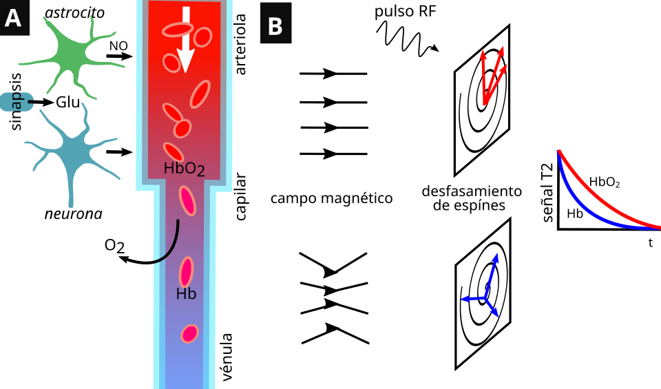
This diagram summarizes how the BOLD signal arises from changes in blood oxygenation following neural activity. It emphasizes that fMRI contrast depends on the balance of oxygenated and deoxygenated hemoglobin, reinforcing that the method measures brain activity indirectly via vascular responses. Source
BOLD signal: Blood-oxygen-level-dependent signal; the change in oxygenated blood that fMRI uses as an indirect measure of neural activity.
This means fMRI does not record neurons firing directly. Instead, it infers brain activity from the blood flow changes that follow neural activity.
How fMRI works
A typical fMRI investigation follows a series of stages:
A participant lies very still inside a scanner while brain images are collected.
The participant may be asked to carry out a task, such as reading words, looking at pictures, or making decisions.
As different brain areas become active, local blood flow and oxygenation change.
The scanner detects these small changes across the brain.
A computer processes the data into detailed images, highlighting areas that show greater activity during the task.
Researchers can then examine whether certain mental processes are consistently linked to activity in particular brain regions. Because the images are highly detailed, fMRI is especially useful for showing where activity occurs in the brain.
fMRI can repeatedly scan the same person over time, which allows changes in activity to be tracked across different conditions within a single study. This makes it valuable for controlled experiments in cognitive neuroscience and psychology.
What fMRI can be used for
In psychology, fMRI is used to investigate the neural basis of behavior and cognition. Researchers can study processes such as:
attention
memory
language
emotion
decision-making
visual processing
It is also useful in clinical contexts. For example, fMRI can help identify brain areas involved in unusual patterns of functioning and may assist with planning treatment or surgery by showing which regions are active during specific functions.
Because fMRI studies the living brain, it has greatly expanded psychologists’ ability to connect mental processes with underlying biological activity. It supports the view that many behaviors and cognitive functions are linked to specialized systems within the brain.
Strengths of fMRI
A major strength of fMRI is that it is non-invasive. It does not require injections, surgery, or exposure to ionizing radiation, so it is considered relatively safe for repeated use in research and clinical practice.
Another important strength is its high spatial resolution. This means it can show activity in very specific brain areas, often down to small regions, allowing researchers to localize function more precisely. This has been especially valuable in building knowledge about which parts of the brain are associated with different cognitive tasks.
fMRI also produces objective data. The images are generated by a scanner and computer analysis rather than relying only on participants’ self-reports. This can increase scientific credibility because the method provides a visible and measurable record of brain activity.
A further advantage is that fMRI can scan the whole brain at once. Researchers are not limited to one small area, so they can investigate networks of activity across several regions during a task.
Limitations of fMRI
Despite its value, fMRI has important limitations. First, it measures brain activity indirectly. The scan shows changes in blood oxygenation, not the actual electrical firing of neurons. As a result, fMRI is an indicator of activity rather than a direct recording of it.
It also has limited accuracy for timing.
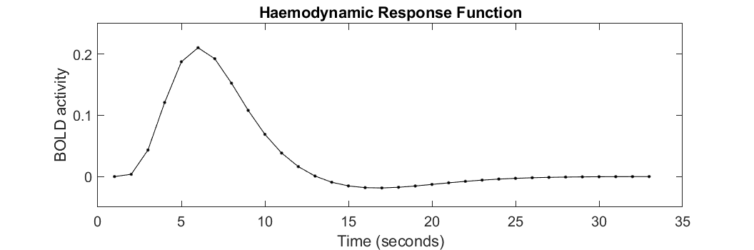
This figure shows a typical hemodynamic response function (HRF)—the time course of the BOLD signal following brief neural activity. The peak occurs several seconds after the underlying neuronal event, illustrating why fMRI has relatively weak temporal precision despite strong spatial localization. Source
Neural firing happens extremely quickly, but the blood flow response detected by fMRI takes longer to develop. This means fMRI is better at showing where activity happens than when it happens with precision.
Another limitation is that fMRI studies are expensive to run. The scanners are costly to buy and maintain, and specialist staff are needed to collect and analyze the data. This can reduce the number of studies carried out and may limit sample sizes.
The method can also be affected by practical issues. Participants must remain very still in the scanner, and even small movements can distort the images. This can make it harder to use with people who are anxious, very young, or unable to keep still for long periods.
Finally, fMRI findings should be interpreted carefully. If a brain area becomes active during a task, this shows an association between that area and the task, but it does not by itself prove that the area alone causes the behavior. Mental processes are often produced by interaction between several brain regions, so oversimplified interpretations can be misleading.
Practice Questions
Outline what fMRI measures in the brain. (2 marks)
1 mark for stating that fMRI measures changes in blood oxygenation or blood flow.
1 mark for linking these changes to neural activity or identifying active brain areas.
Discuss one strength and one limitation of fMRI as a way of studying the brain. (6 marks)
1 mark for identifying a clear strength, such as being non-invasive, objective, or having high spatial resolution.
1 mark for explaining that strength.
1 mark for showing why that strength is useful in psychological research.
1 mark for identifying a clear limitation, such as being an indirect measure, expensive, or sensitive to movement.
1 mark for explaining that limitation.
1 mark for showing why that limitation reduces usefulness or affects interpretation of findings.
FAQ
A voxel is a tiny three-dimensional unit of brain image data, similar to a pixel but with volume.
Smaller voxels can give more detailed information about where activity is located, but they also produce weaker signals and may require longer scanning or more complex analysis.
Larger voxels are easier to detect reliably, but they blur together activity from a wider area. This means voxel size affects how precisely researchers can map brain activity.
Yes. This is called resting-state fMRI.
Instead of asking the participant to complete a task, researchers scan the brain while the person rests quietly. They then look for patterns of activity that rise and fall together across different regions.
This can help identify large-scale brain networks that seem to work together even when no specific task is being performed.
fMRI uses a very strong magnetic field, so metal can create serious safety risks or distort the image.
Problems include:
movement or heating of some metal objects
interference with medical devices such as pacemakers
image artifacts that make results hard to interpret
Because of this, participants are carefully screened before scanning. Not all metal is unsafe, but it must always be checked first.
Raw fMRI data usually goes through several preprocessing steps before analysis.
These often include:
correcting for small head movements
aligning each brain image to a standard brain space
smoothing the data to reduce noise
removing obvious distortions or scanner errors
These steps help make results more reliable and allow researchers to compare participants more fairly. However, preprocessing choices can also influence the final pattern of results.
Individual brains differ in size, shape, and exact functional layout, so one person’s scan may not represent the general population well.
By combining data from many participants, researchers can:
reduce the effect of unusual individual patterns
improve reliability
identify activity that appears consistently across people
This makes it easier to draw broader conclusions. However, averaging can also hide important individual differences, especially in areas such as language or recovery after brain injury.
