HL only: topic status
· C2.1 Chemical signalling is entirely HL content (no SL content).
· Focus on how cells detect ligands, how receptors trigger intracellular responses, and how signalling pathways are regulated.
· Exam answers should always link ligand → receptor → transduction pathway → cellular response.
Receptors and ligand specificity
· Receptors are proteins with specific binding sites for a ligand (signalling chemical).
· Specificity depends on the 3D shape and chemical properties of the binding site.
· Only cells with the correct receptor can respond to a particular signal.
· Ligand binding causes a conformational change in the receptor, starting a signal transduction pathway.
· A signalling pathway is a sequence of intracellular events that converts an external signal into a cellular response.
Quorum sensing in bacteria
· Quorum sensing = cell signalling in bacteria based on population density.
· Bacteria release small signalling molecules; as population size rises, their concentration rises.
· When a threshold concentration is reached, many cells change gene expression simultaneously.
· In Vibrio fischeri, quorum sensing switches on bioluminescence.
· This allows a coordinated response across the population rather than isolated responses by single cells.
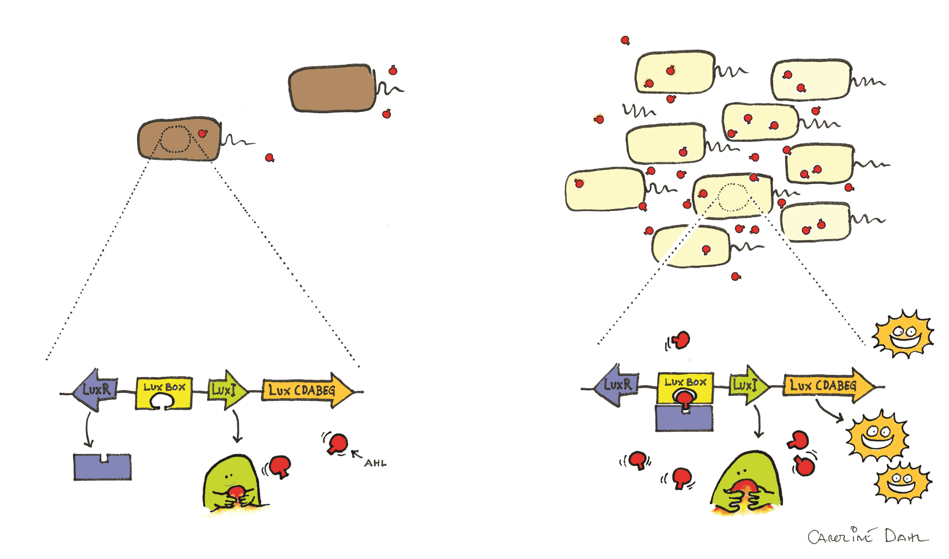
This diagram shows how autoinducer molecules accumulate as bacterial population density increases, eventually activating genes for light production in Vibrio fischeri. It is useful for linking ligand concentration to a coordinated gene expression response. Source
Functional categories of signalling chemicals in animals
· Hormones: chemical signals released by endocrine glands and usually transported in the blood to distant target cells.
· Neurotransmitters: chemicals released by neurons that diffuse across a synaptic cleft to a nearby target cell.
· Cytokines: signalling proteins used especially in cell communication in the immune system.
· Calcium ions (Ca2+) can act as intracellular signalling molecules.
· Exam contrast: hormones often act over long distances, whereas neurotransmitters act locally and rapidly.
Chemical diversity of signalling molecules
· A wide variety of chemicals can act as signals because different functions require different solubilities, transport methods, speeds of action and target locations.
· Hormones may be amines, proteins/peptides, or steroids.
· Neurotransmitters may be amino acids, peptides, amines, or nitric oxide.
· Protein/peptide hormones are generally hydrophilic and bind to cell-surface receptors.
· Steroid hormones are hydrophobic, cross membranes, and bind intracellular receptors.
Local vs distant signalling
· Some signalling is localized, affecting nearby cells only.
· Some signalling is distant, with signals transported to far-away target cells.
· Neurotransmitters usually cross only a tiny synaptic gap.
· Hormones are usually carried in the bloodstream to target cells elsewhere in the body.
· Exam tip: always relate distance travelled to transport method and speed/specificity of response.
Transmembrane vs intracellular receptors
· Transmembrane receptors are embedded in the plasma membrane.
· Their outer region binds an external ligand; the inner region triggers intracellular changes.
· They are suited to hydrophilic ligands, which cannot cross the phospholipid bilayer easily.
· Intracellular receptors are in the cytoplasm or nucleus.
· They bind hydrophobic ligands that can pass through the membrane, such as steroid hormones.
· In membrane receptors, amino acids exposed to the membrane are often hydrophobic; regions exposed to aqueous environments are more hydrophilic.
Neurotransmitter receptors and membrane potential
· Some transmembrane receptors are ligand-gated ion channels.
· Example: the acetylcholine receptor.
· When acetylcholine binds, the receptor opens an ion channel.
· Positive ions diffuse into the cell, changing the membrane potential.
· This can trigger further cellular responses, including depolarization.
· Key idea: signal conversion here is very direct — ligand binding immediately changes ion movement across the membrane.
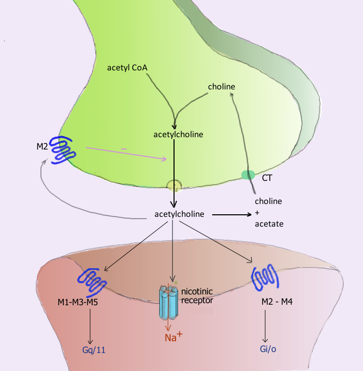
This image shows release of acetylcholine, diffusion across the synaptic cleft, and binding to receptors on the postsynaptic membrane. It helps explain how neurotransmitter binding can rapidly change membrane potential. Source
G protein-coupled receptors (GPCRs)
· Some transmembrane receptors activate G proteins on the inside of the membrane.
· A ligand binds to the receptor, changing its shape and activating the G protein.
· The activated G protein then triggers another enzyme or protein in the membrane.
· This starts an intracellular signal cascade.
· GPCRs are extremely common in humans and are used in many signalling systems.
Adrenaline (epinephrine) and cAMP
· Epinephrine (adrenaline) acts through a G protein-coupled receptor.
· Ligand binding activates a G protein.
· The G protein activates adenylyl cyclase.
· Adenylyl cyclase converts ATP into cyclic AMP (cAMP).
· cAMP acts as a second messenger inside the cell.
· Second messenger = intracellular signalling molecule that relays the message from the membrane receptor.
· The pathway amplifies the signal, so one ligand-binding event can cause a large cellular response.
· In exams, state clearly: adrenaline is the first messenger; cAMP is the second messenger.
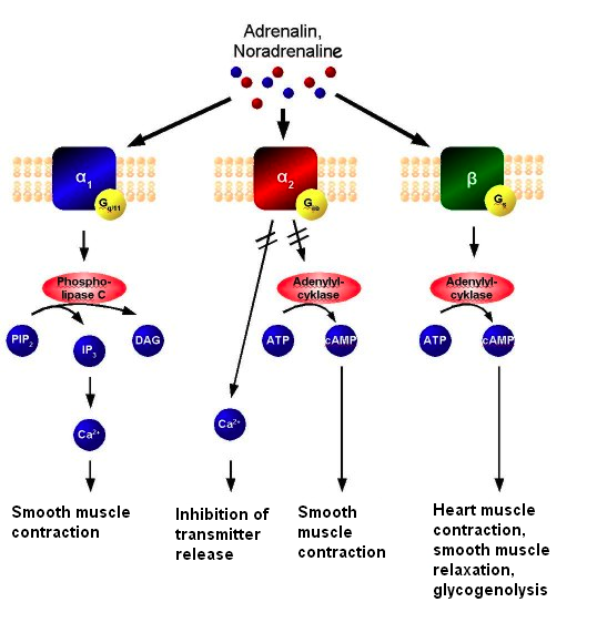
This diagram shows how adrenaline binds to a GPCR, activates a G protein, and leads to cAMP-mediated intracellular signalling. It is especially useful for revising the idea of a second messenger. Source
Receptors with tyrosine kinase activity: insulin
· Some transmembrane receptors have tyrosine kinase activity.
· Example: the insulin receptor.
· Insulin binds to the receptor on the plasma membrane.
· This causes phosphorylation of tyrosine on the intracellular part of the receptor.
· A signalling cascade follows inside the cell.
· One outcome is movement of vesicles containing glucose transporters to the plasma membrane.
· This increases glucose uptake by the cell.
· Exam wording: insulin binding leads to receptor activation → phosphorylation → intracellular cascade → insertion of glucose transporters into the membrane.
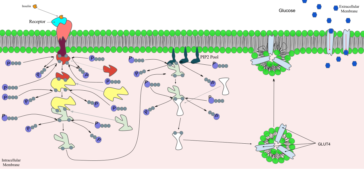
This diagram shows how insulin binding triggers phosphorylation and a signalling cascade that changes cell activity. It supports the syllabus idea that insulin signalling ends with glucose transporter vesicles moving to the plasma membrane. Source
Intracellular receptors and gene expression
· Intracellular receptors are activated by hydrophobic ligands that can pass through the plasma membrane.
· Example ligands: oestradiol, progesterone and testosterone.
· The hormone binds its receptor in the cytoplasm or nucleus.
· This activates the receptor.
· The activated receptor binds to specific DNA sequences.
· This promotes gene transcription and changes protein synthesis in the target cell.
· Responses are often slower than membrane-receptor pathways because they depend on changes in gene expression.
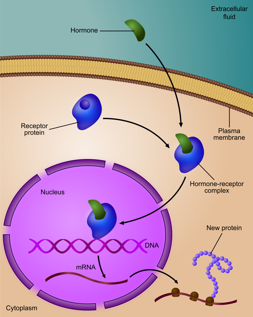
This image shows a steroid hormone entering the cell, binding an intracellular receptor, and the activated complex interacting with DNA to alter transcription. It is ideal for comparing intracellular receptors with cell-surface receptors. Source
Effects of oestradiol and progesterone
· Oestradiol acts on cells in the hypothalamus that secrete gonadotropin-releasing hormone (GnRH).
· Progesterone acts on cells in the endometrium.
· Because these are steroid hormones, they act through intracellular receptors that alter gene expression.
· Exam tip: link each hormone to its target tissue/cells and to the general mechanism of intracellular receptor activation.
Feedback regulation of signalling pathways
· Negative feedback reduces or counteracts the original change.
· It helps maintain stability and prevents over-response.
· Positive feedback reinforces the original change.
· It drives a process forward more strongly once it has started.
· In signalling pathways, feedback can regulate hormone release, pathway activity, or cellular sensitivity.
· In exam questions, be ready to distinguish clearly between stabilizing (negative) and amplifying (positive) feedback.
Checklist: can you do this?
· Explain why only target cells with the correct receptor respond to a ligand.
· Compare transmembrane and intracellular receptors using membrane permeability and ligand type.
· Describe the pathway for adrenaline via GPCR → G protein → adenylyl cyclase → cAMP.
· Explain how insulin signalling leads to glucose transporter vesicles moving to the membrane.
· Interpret an unfamiliar diagram of a signalling pathway by identifying ligand, receptor, second messenger, and response.
High-yield exam links
· Hydrophilic ligand → usually cell-surface receptor.
· Hydrophobic ligand → usually intracellular receptor.
· Neurotransmitter receptor may change membrane potential very rapidly.
· GPCR pathways often use second messengers and signal amplification.
· Steroid hormones often act by changing transcription.
· Always write answers as a cause-and-effect chain rather than isolated facts.