Neurons: structure and role
Neurons are cells of the nervous system that carry electrical impulses.
Cell body contains the cytoplasm and nucleus.
Dendrites are short, branched fibres that receive signals.
Axon is a single long fibre that conducts impulses away from the cell body.
Signals move as electrical changes across the plasma membrane.
At the end of neurons, signals can be passed to other neurons or to effector cells such as muscle cells.
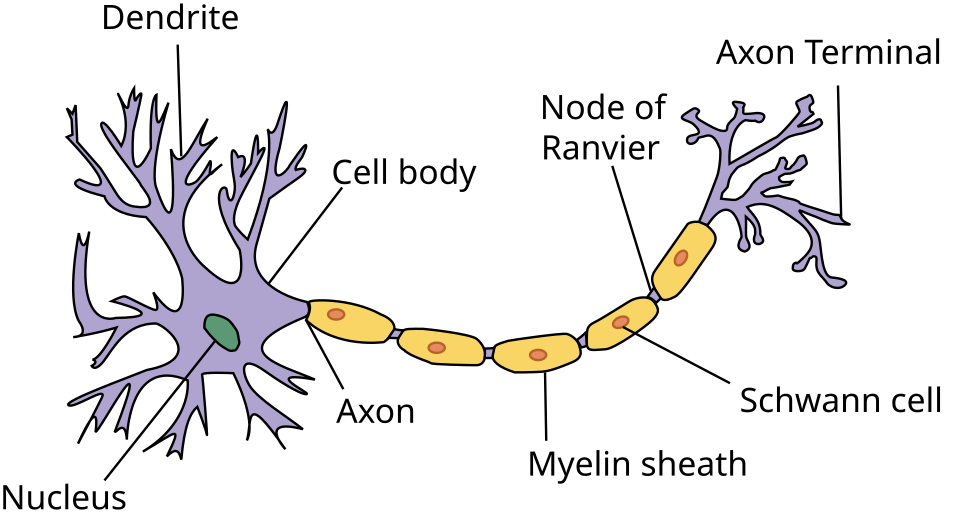
This diagram shows the basic structure of a neuron, including dendrites, cell body, axon, myelin sheath, and nodes of Ranvier. It is useful for identifying the pathway along which impulses travel. Source
Resting potential
A neuron at rest has a resting potential: the membrane is polarized.
This is generated by active transport using the sodium–potassium pump.
The pump moves sodium ions (Na⁺) out and potassium ions (K⁺) in using ATP.
This establishes concentration gradients for both ions across the membrane.
The inside of the neuron is negative relative to the outside.
Key idea: membrane potential = voltage across the membrane; polarization = charge difference across the membrane.
Action potentials and nerve impulses
A nerve impulse is an action potential propagated along a nerve fibre.
It is electrical because it involves movement of positively charged ions.
In HL, you must know the stages:
Threshold potential must be reached.
Voltage-gated sodium channels open → Na⁺ enters.
Membrane becomes less negative then positive = depolarization.
Voltage-gated sodium channels close and voltage-gated potassium channels open.
K⁺ leaves the axon = repolarization.
Membrane may briefly become more negative than resting = hyperpolarization.
All-or-nothing principle: if threshold is not reached, no action potential is produced.
After an action potential, there is a refractory period, helping ensure one-way transmission.
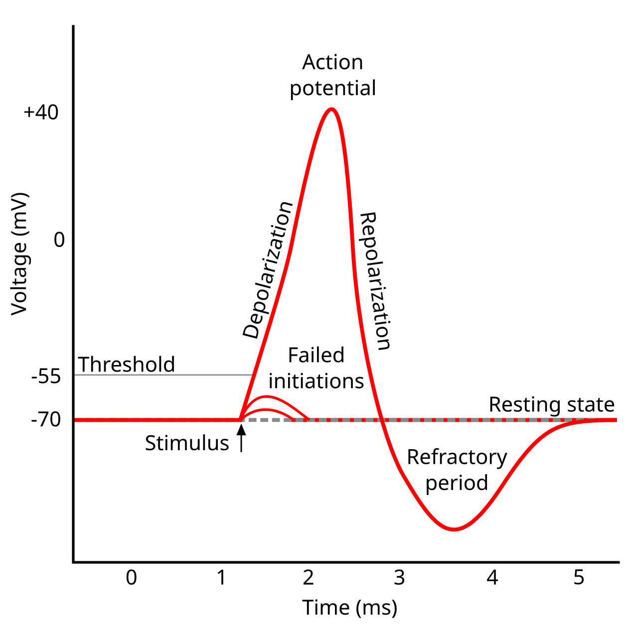
This graph shows the voltage changes during an action potential: resting state, threshold, depolarization, repolarization, and refractory period. It is ideal for linking membrane events to a trace in exam questions. Source
Propagation along the axon
Action potentials move along the axon due to local currents.
Entry of Na⁺ at one region causes diffusion of positive charge to adjacent regions.
This brings the next section of membrane to threshold, so a new action potential is generated there.
Therefore the impulse is propagated along the fibre.
Impulses travel faster in larger diameter axons because internal resistance is lower.
Compare:
Giant squid axons conduct rapidly because of large diameter.
Small non-myelinated fibres conduct more slowly.
Be able to interpret correlations:
Conduction speed is positively correlated with axon diameter.
Conduction speed is negatively correlated with animal size in the syllabus example.
Myelination and saltatory conduction
Myelin sheath insulates the axon.
Gaps in myelin are nodes of Ranvier.
Ion channels and ion pumps are concentrated at the nodes.
In myelinated axons, action potentials occur only at the nodes.
The impulse effectively jumps from node to node = saltatory conduction.
This makes transmission much faster than in non-myelinated fibres.
Myelination also reduces energy use because fewer membrane regions need ion pumping.
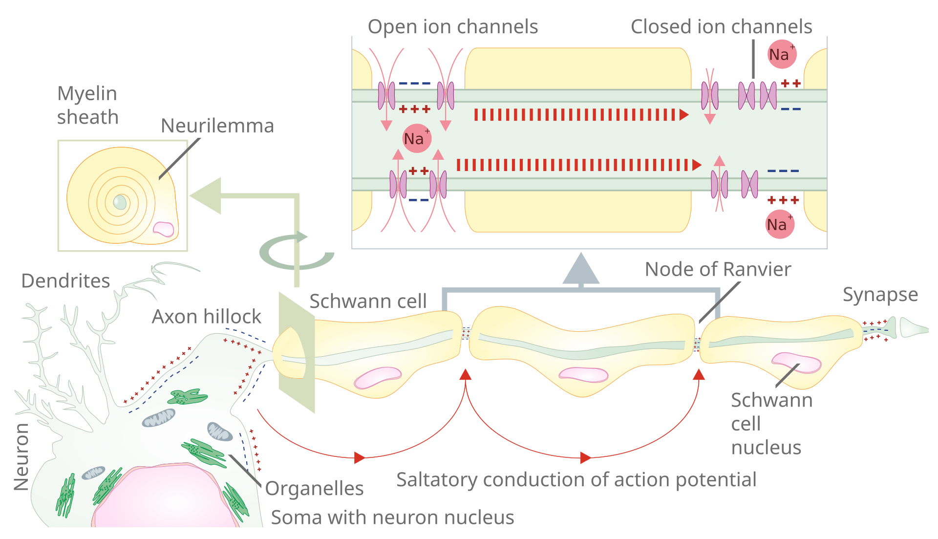
This diagram shows saltatory conduction in a myelinated axon, with action potentials moving between nodes of Ranvier. It helps explain why myelinated fibres conduct impulses faster than non-myelinated fibres. Source
Synapses and synaptic transmission
A synapse is a junction between neurons or between a neuron and an effector cell.
In IB Biology, focus on chemical synapses.
Transmission across a synapse is one-way.
Sequence of synaptic transmission:
Action potential reaches the presynaptic membrane.
Depolarization opens calcium ion channels.
Ca²⁺ enters the presynaptic neuron.
Calcium acts as an internal signalling chemical.
Vesicles fuse with the presynaptic membrane and release neurotransmitter.
Neurotransmitter diffuses across the synaptic cleft.
It binds to transmembrane receptors on the postsynaptic membrane.
Acetylcholine is the named example you should know.
At many synapses, receptor binding opens ion channels and changes the postsynaptic membrane potential.
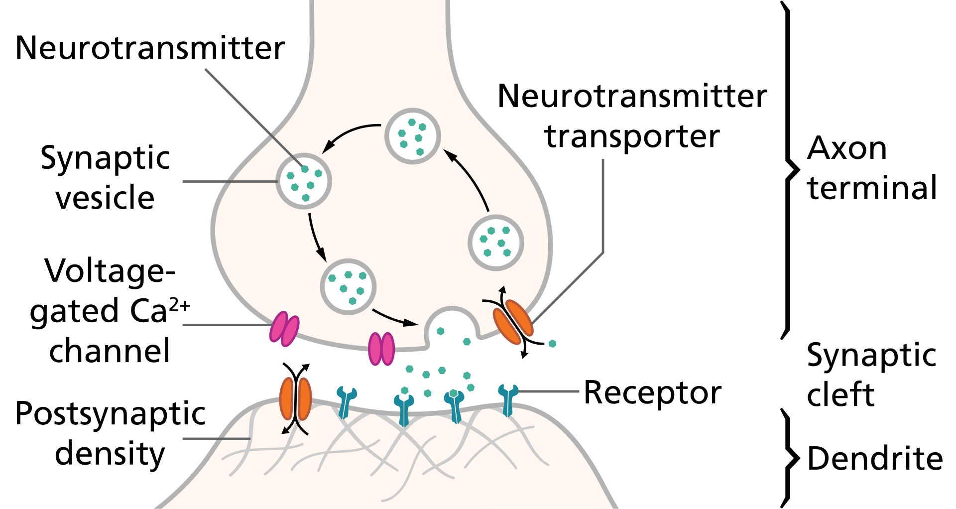
This figure shows vesicles releasing neurotransmitter, diffusion across the synaptic cleft, and binding to postsynaptic receptors. It is helpful for explaining why synaptic transmission is one-way. Source
Excitatory and inhibitory postsynaptic potentials
An excitatory postsynaptic potential (EPSP) makes the postsynaptic membrane less negative.
Example: acetylcholine binds receptors, allowing positive ions to enter.
If threshold is reached, a new action potential is generated in the postsynaptic neuron.
An inhibitory postsynaptic potential (IPSP) makes the postsynaptic membrane more negative = hyperpolarization.
Inhibitory neurotransmitters therefore make an action potential less likely.
The postsynaptic neuron sums all inputs:
Excitatory inputs push toward threshold.
Inhibitory inputs move away from threshold.
This is summation and leads to all-or-nothing output in the postsynaptic neuron.
Speed, data, and graph interpretation
Be able to compare myelinated vs non-myelinated fibres.
Be able to compare large-diameter vs small-diameter axons.
Know the difference between:
Positive correlation: both variables increase together.
Negative correlation: one variable increases as the other decreases.
Correlation coefficient shows the strength and direction of a relationship.
Coefficient of determination (R²) shows how much variation in the dependent variable is explained by the independent variable.
On an oscilloscope trace, relate shape to events:
flat negative line = resting potential
steep rise = depolarization
steep fall = repolarization
undershoot = hyperpolarization / refractory period
Frequency of impulses can be measured as number of action potentials per second.
HL only: action potential detail and oscilloscopes
Know the roles of voltage-gated sodium channels and voltage-gated potassium channels.
Threshold potential must be reached before sodium channels open.
Local currents explain how one action potential triggers the next region of membrane.
Be able to interpret oscilloscope traces and connect them to ion channel activity.
Resting potential, depolarization, repolarization, and hyperpolarization should be recognized on graphs.
HL only: synaptic modulation and drugs
Exogenous chemicals can alter synaptic transmission.
Neonicotinoids are pesticides that block synaptic transmission.
Cocaine blocks reuptake of neurotransmitter, so neurotransmitter remains longer in the synaptic cleft.
This prolongs or alters signalling at the synapse.
Questions may ask how blocking reuptake or receptors changes postsynaptic stimulation.
HL only: pain perception and emergence
Pain receptors in skin are neurons with free nerve endings.
Their ion channels open in response to stimuli such as high temperature, acid, or capsaicin.
Entry of positive ions causes the membrane to reach threshold.
Action potentials then pass to the brain, where pain is perceived.
Consciousness is treated as an emergent property arising from interactions among many neurons in the brain.
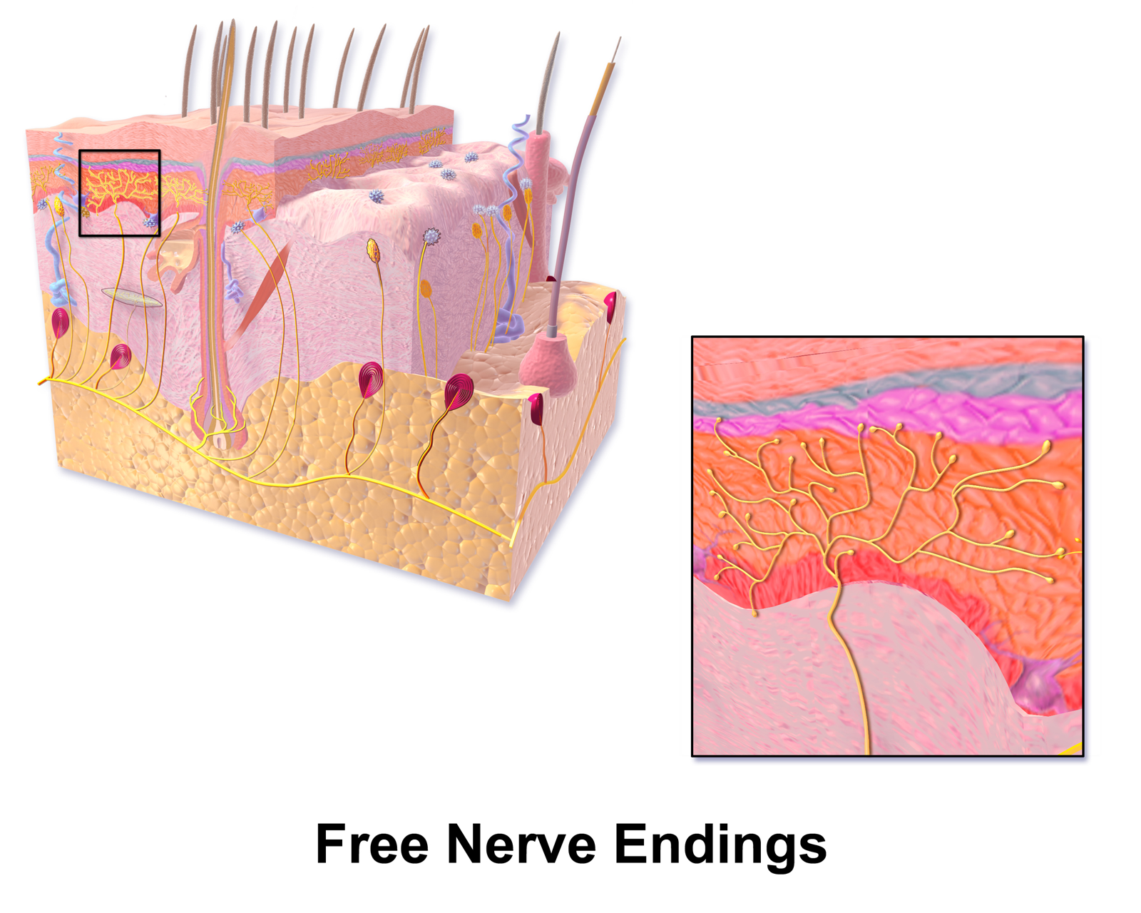
This diagram shows free nerve endings in the skin, the type of sensory ending linked to pain detection in the syllabus. It helps connect external stimuli to ion channel opening and action potential generation. Source
Checklist: can you do this?
Label a neuron and distinguish cell body, dendrites, axon, myelin sheath, and nodes of Ranvier.
Explain how the resting potential is generated by the sodium–potassium pump and why it is negative.
Describe and interpret the stages of an action potential, including threshold, depolarization, repolarization, and refractory period.
Explain how transmission across a chemical synapse occurs and why it is one-way.
Interpret data or graphs on conduction speed, correlation, R², and oscilloscope traces.
Exam traps and quick fixes
Do not say impulses are the movement of electrons; they involve movement of ions.
Do not confuse resting potential with action potential.
Do not say neurotransmitter crosses by active transport; it crosses the synaptic cleft by diffusion.
Do not forget that Ca²⁺ entry into the presynaptic neuron triggers neurotransmitter release.
Do not confuse EPSP with an actual action potential; an EPSP is a small depolarization that may help reach threshold.
When comparing speed, always mention axon diameter and myelination.
In HL, remember that threshold must be reached before voltage-gated sodium channels open.
Ultra-compact memory lines
Resting potential = Na⁺ out, K⁺ in, inside negative.
Action potential = threshold → Na⁺ in → K⁺ out.
Myelin = faster conduction by jumping between nodes.
Synapse = Ca²⁺ in → neurotransmitter out → receptors bind.
EPSP pushes toward threshold; IPSP pushes away from th